Reconstructed Human Epidermis (RHE) – ISO 10993-23, OECD 439
The in vitro Skin Irritation Test is an OECD-approved in vitro non-animal test method for identifying Test Materials that may be irritating to reconstructed human epidermis.
The in vitro skin irritation test is used for the hazard identification of irritant chemicals in accordance with the UN Globally harmonized system of classification and labelling (GHS) Category 2. It aims to predict test compounds potential to cause dermal irritation.
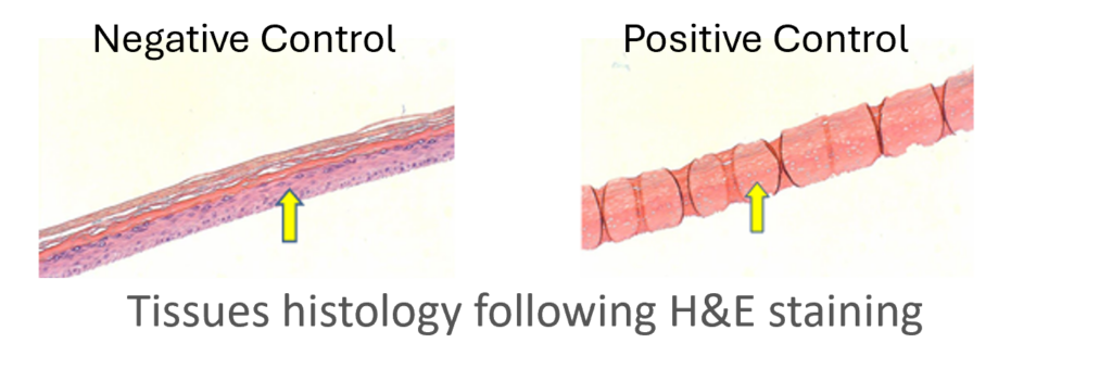
The reconstructed human epidermal tissues consists of normal human-derived epidermal keratinocytes, which have been cultured to form a multi-layered highly differentiated model of the human epidermis. It consists of organized basal, spinous and granular layers, and a multi-layered stratum corneum containing intercellular lamellar lipid layers arranged in patterns analogous to those found in vivo.
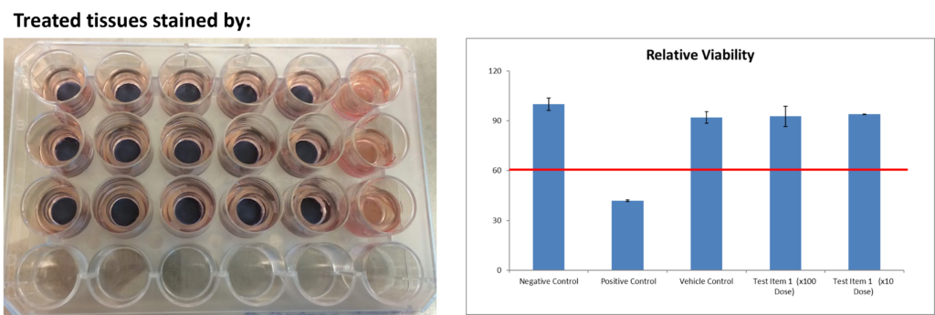
The test consists of exposure of the test materials to the reconstructed human epidermis construct tissues followed by a cell viability test using MTT.
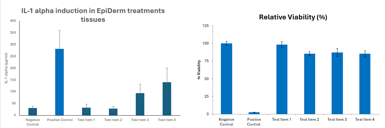
MTT assay has been shown to generally be sufficient for predicting irritation, however, various implementations of IL-1α as a secondary endpoint have been used in the industry. These include using IL-1α results to verify borderline MTT assay results or using IL-1α to verify all non-irritant results in the MTT assay.
The results are reported as percent viability (% of living cells).
Test materials are considered non irritant when cells viability is ≥ 50%.
In vitro Eye Irritation Test:
Reconstructed human Cornea-like Epithelium (RhCE) – OECD 492 , ISO 10993-23
The in vitro Eye Irritation Test is an OECD-approved in vitro non-animal test method for identifying Test Materials that may be irritating to reconstructed human corneal epithelium.
The methods is used as an alternative to the traditional In vivo – rabbit eye test method (OECD TG 405).
The test consists of a topical exposure of Test Material onto the Human Corneal Epithelium model HCE, followed by cell viability measurement.
The Reconstructed human cornea-like epithelium is a three-dimensional non-keratinized tissue construct composed of normal human derived epidermal keratinocytes. It consists of highly organized basal cells which progressively flatten out as the apical surface of the tissue and is analogous to the normal in vivo corneal epithelium. The tissue is mitotically and metabolically active and releases many of the pro-inflammatory agents (cytokines) known to be important in ocular irritation and inflammation.
The test consists of exposure of the test materials to the reconstructed human cornea epithelial construct tissues followed by a cell viability test using MTT.
The results are reported as percent viability (% of living cells).
Test materials are considered non irritant when cells viability is ≥ 60%.
HET-CAM assay:
ICCVAM-recommended test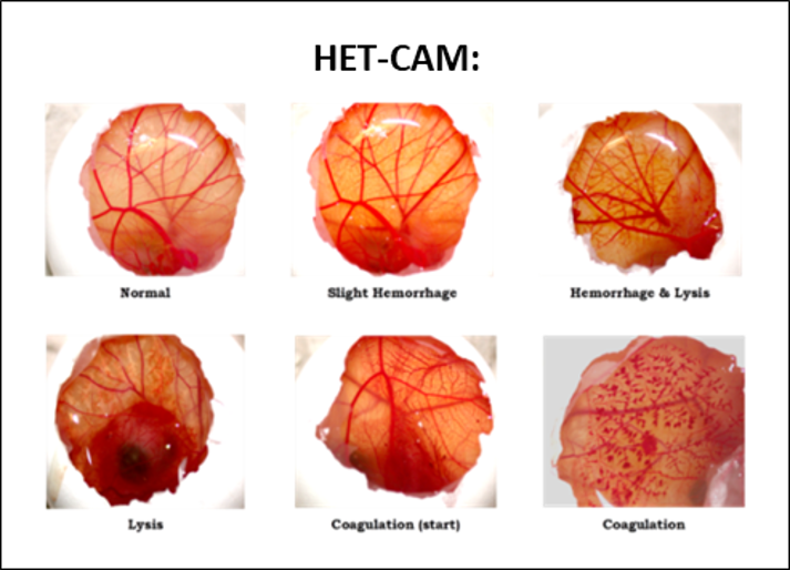 method
The Hen’s Egg Test – Chorioallantoic Membrane (HET-CAM) is used to determine the potential irritancy of chemicals, medical devices and materials that are in contact with the eyes or mucous membranes. It can also be suitable for textiles, cosmetics and water.
The HET-CAM is a model that is proposed to mimic the mucosal tissues of the eye and detects vascular injury.
The potential of Test Material to cause irreversible or severe eye irritation or corrosion may be detected by observing adverse changes, which occur in the egg’s CAM after exposure to the test material.
The CAM is a complete tissue containing arteries, veins and capillaries, and is technically easy to study. It responds to injury with an inflammatory process similar to what can be observed in the conjunctival tissue of a rabbit’s eye.
Its well-developed vascularization provides an ideal model for ocular irritation studies.
Test Material is extracted or applied as-is and placed on the egg’s CAM for a few minutes and he blood vessels coagulation, hemorrhage, and lysis are determined.
GMP Batch Release Testing is conducted for every manufactured drug product batch to ensure each batch is safe and effective for use by patients before it is released to the market.
Cytokine evaluation:
Detecting and understanding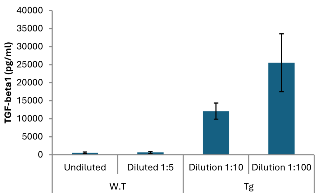 of the cytokine secretions provides vast information on mechanisms, treatments and the development of new treatments.
Cytokine detection has been used as an inflammatory biomarker for the diagnosis of many diseases.
Cytokines profiles can be characterized in cancer disease, cytokine storm, autoimmune disease, inflammation cytokines, apoptosis etc.
A single cytokine can be detected by ELISA while panels of cytokines can be simultaneously detected by multiplex platforms such as Luminex and flow cytometry.
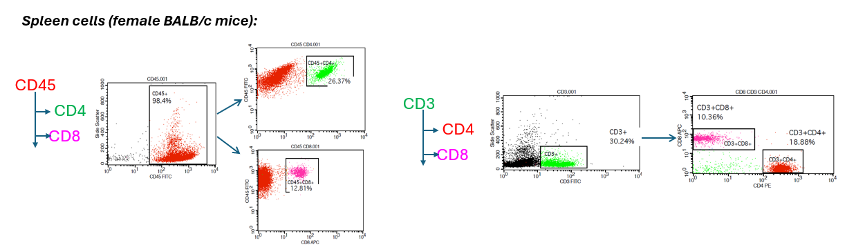
The power of flow cytometry is to provide insights into cell populations.
Characterization of various immune cells subtypes by virtue of size and morphology: T cell, B cell, monocyte, NK cell etc.
HBI Services offers bioanalysis using Flow Cytometry for preclinical development and our team help you to establish the most appropriate assay for your research.
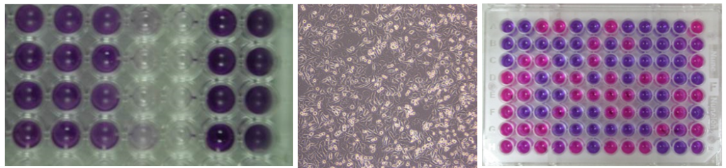
MTT
XTT
Alamar BlueTM
 The development of monoclonal antibodies (mAbs) remains high on the therapeutic agenda for the majority of pharmaceutical and biotechnology companies.
The mAbs produced have broad utility as therapeutics, diagnostic tools, and research reagents.
HBI is an expert in the usage of mAbs development technology.
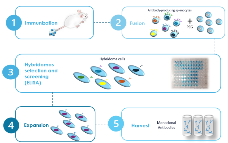
mAbs technology is based on immunization of mice with a specific antigen to prompt a humoral immune response. Antibody-producing B-cells cells from the most immune responder mouse, are fused with a myeloma cell line to generate hybrid cells that are sequentially cloned to obtain stable monoclonal hybridoma.
MAbs resemble natural immunoglobulins from the immunized mouse but differ from serum-derived polyclonal antibodies as they are specific to a single epitope, ensuring a stable, long-term supply.
The MAbs manufacturing process plays an important role in producing therapeutic antibodies widely used in treating diseases such as cancer, autoimmune disorders and other infectious diseases.
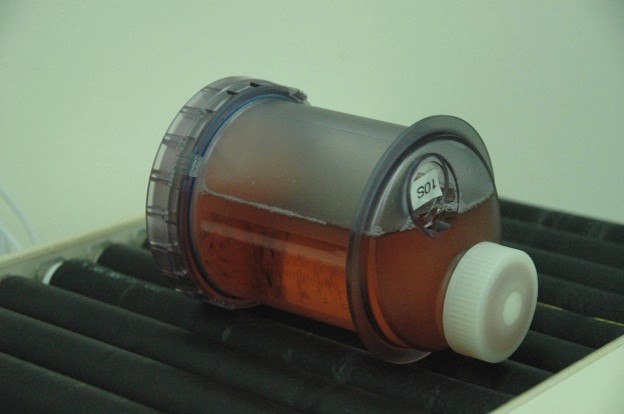
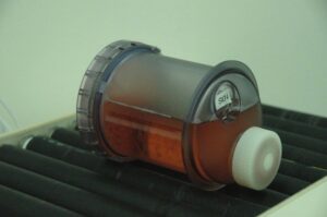
HBI offer mAbs production services using bioreactors technology.